Scanning Transmission Electron Microscopy (STEM) Detector
![Scanning Transmission Electron Microscopy coxem](https://admin.jhtechnologies.com/sites/default/files/styles/1000/public/image%20%285%29Scanning%20Transmission%20Electron%20Microscopy.jpg?itok=WNccziCR)
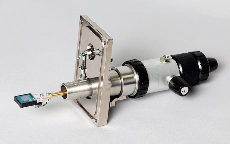
Our STEM Detector offers the ability to use either the EM-30 Series Tabletop SEM or the CX-200plus SEM to perform STEM analysis with standard TEM grids.
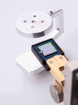
The STEM detector mounts to the SEM via a side port. When not used, the STEM detector retracts and tilts 90° out of view for standard SEM analysis. The detector can collect both Bright Field (BF) and Dark Field (DF) images.
- Bright Field (BF) and Dark Field (DF) imaging
- Retractable Detector
- Detector position adjustable
- Sample Holder for 4 TEM grids included
- Stage X-Y used to position any of 4 sample locations
- Dedicated Detector Image channel in NS 4.0 software
![COXEM STEM Module for SEM hardware](https://admin.jhtechnologies.com/sites/default/files/styles/1000/public/image%20%285%29.jpg?itok=DrEz51yr)
Example STEM Images
![Mouse Kidney Magnification](https://admin.jhtechnologies.com/sites/default/files/styles/1000/public/image%20%286%29.jpg?itok=BdM4001p)
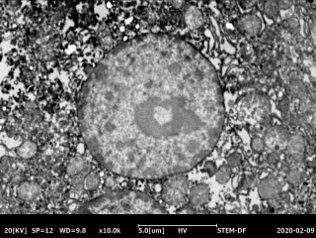
Mouse Kidney at 10,000 Magnification
Learn More About (STEM) Detector
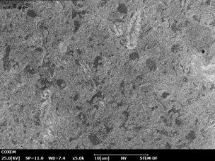
![Neuron Cells from 5,000 STEM](https://admin.jhtechnologies.com/sites/default/files/styles/1000/public/image%20%287%29.jpg?itok=KW4Kwmfa)
![Neuron Cells from 30,000X STEM](https://admin.jhtechnologies.com/sites/default/files/styles/1000/public/image%20%288%29.jpg?itok=q8LP09wh)
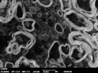
Neuron Cells from 5,000 to 30,000X Magnification
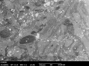
![Colon Cancer cells at 3,000 STEM](https://admin.jhtechnologies.com/sites/default/files/styles/1000/public/Colon%20Cancer%20cells%20at%203%2C000.png?itok=kRdR0DqB)
![Colon Cancer cells at 30,000X Magnification STEM](https://admin.jhtechnologies.com/sites/default/files/styles/1000/public/Colon%20Cancer%20cells%20at%2030%2C000X%20Magnification.jpg?itok=R_DnPZNH)
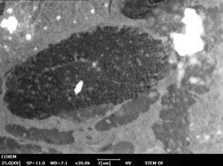
Colon Cancer cells at 3,000 to 30,000X Magnification: